 Module 1 · Classification
Module 1 · Classification
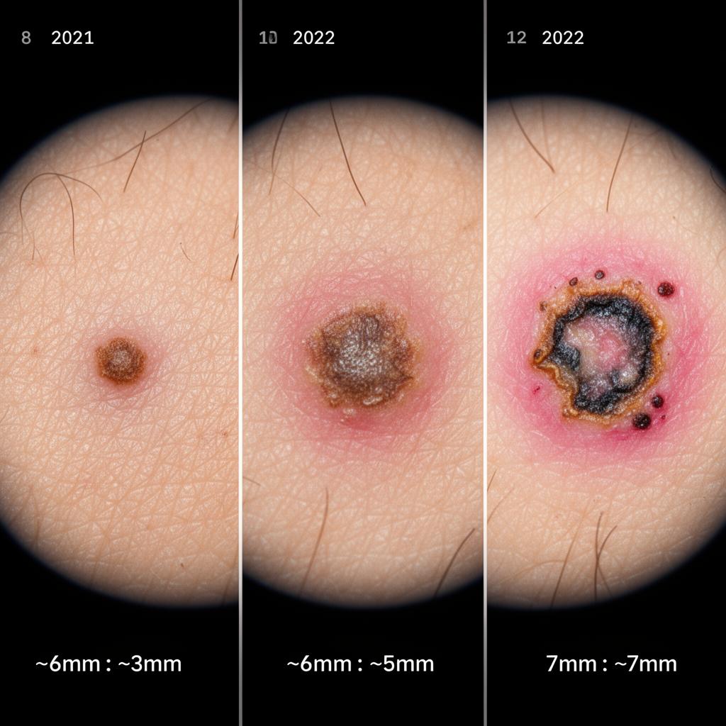
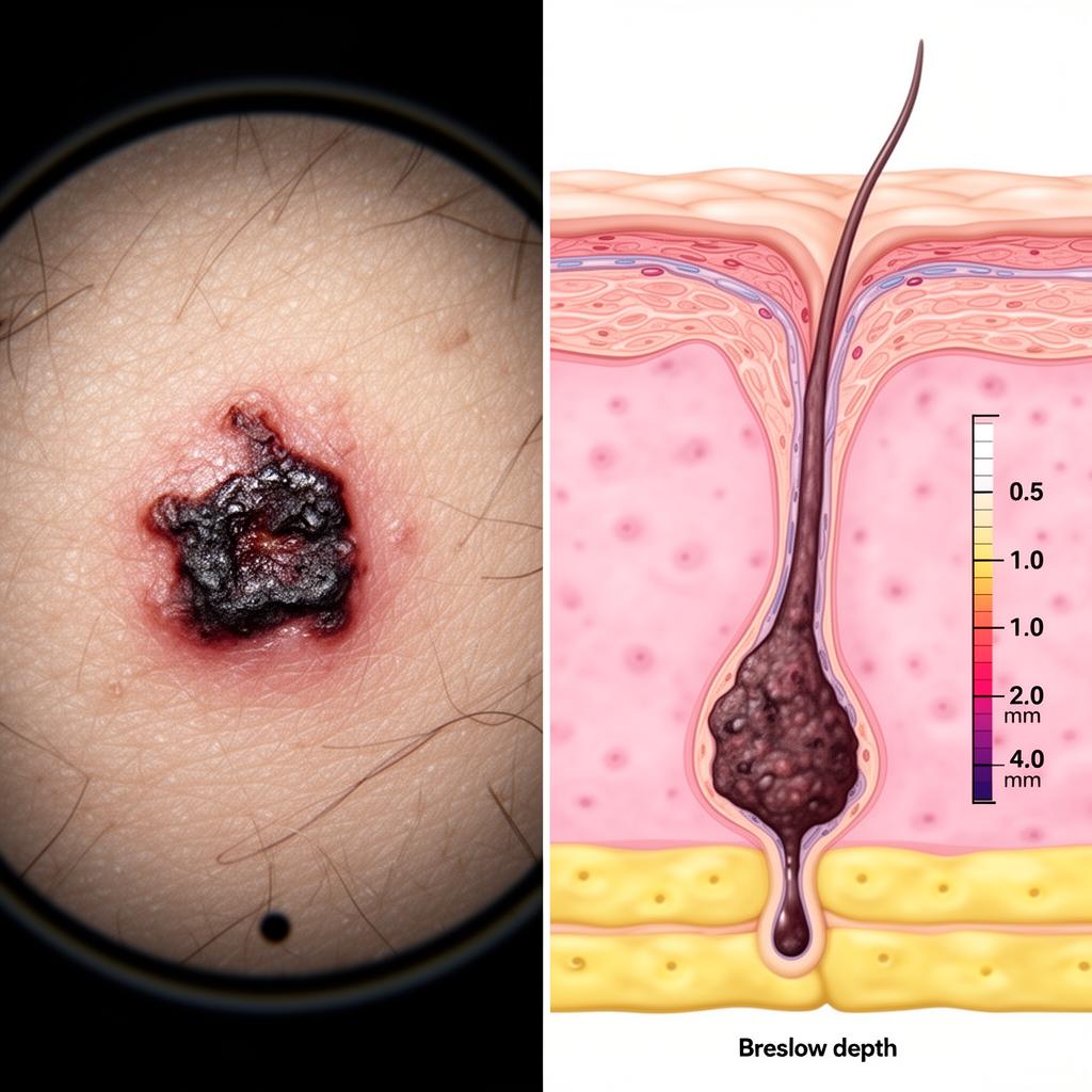
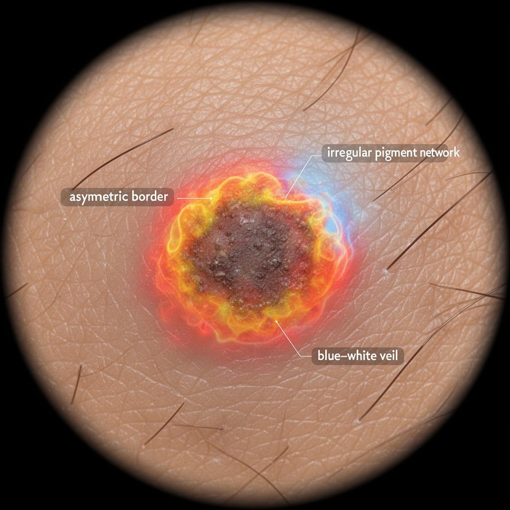
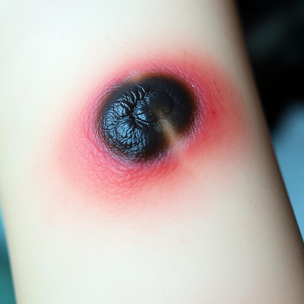
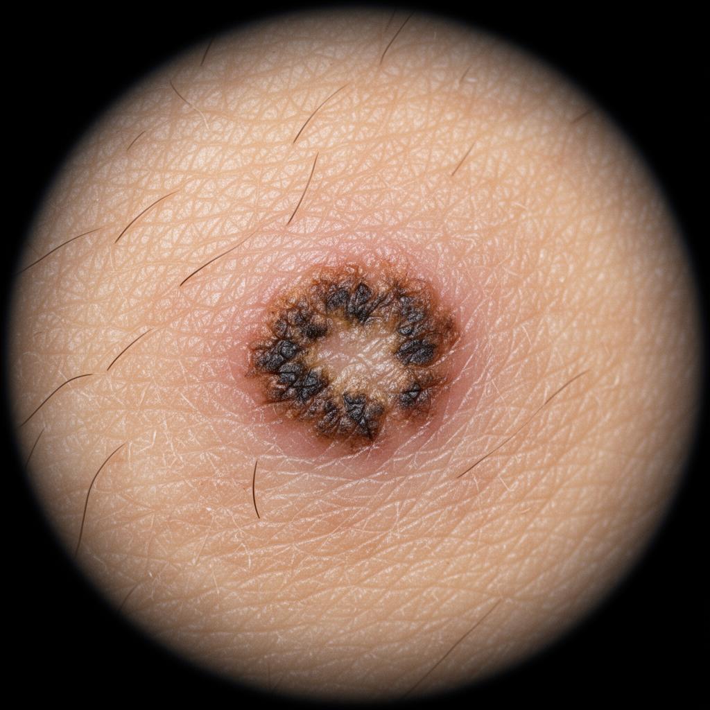
Lesion Classification
An 8-class dermatology taxonomy that distinguishes melanoma, basal cell carcinoma, squamous cell carcinoma, actinic keratosis, benign nevi, seborrheic keratosis, vascular lesions and dermatofibromas — with calibrated confidence and a top-3 differential.
- Works on both dermoscopic and smartphone images (140+ skin conditions covered)
- Top-3 differential diagnoses with probability bars
- Temperature-calibrated confidence — no over-confident outputs
- Built-in 'not skin' detector rejects non-clinical photos
95%
Accuracy · ISIC validation set
8 + 140
Cancer classes · skin conditions